Introduction to Fluorescence Microscopy
Fluorescence microscopy is a cornerstone of biomedical research, enabling detailed visualization of cellular structures. Optical filters are critical components, ensuring precise wavelength control for clear, high-contrast images.
How Fluorescence Microscopy Works
A fluorescence microscope uses a high-energy light source to excite a sample. The light passes through an excitation filter, selecting a specific wavelength, and is reflected by a dichroic mirror onto the sample. The sample emits fluorescence, which passes through the dichroic mirror and an emission filter before being observed by the eye or captured by a camera.
Role of Optical Filters
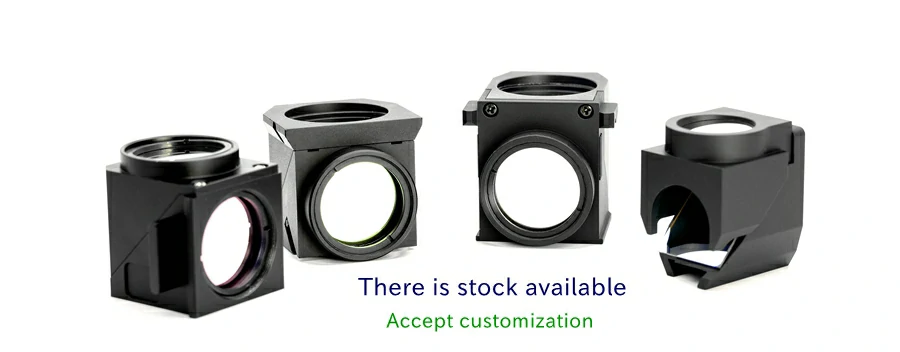
The filter system, comprising the excitation filter, dichroic mirror, and emission filter, is essential for image quality:
- Excitation Filter: Selects the wavelength to excite the sample’s fluorophores.
- Dichroic Mirror: Separates excitation and emission light.
- Emission Filter: Blocks excitation light with high optical density, ensuring only the weaker fluorescence signal is captured, enhancing clarity.
Choosing the Right Filters
Filter selection depends on the sample’s excitation and emission wavelengths. For non-fluorescent samples, fluorescent probes are used to enable imaging. Precise filter matching ensures high signal-to-noise ratios, critical for applications like cellular analysis or disease research.
Why Choose Our Fluorescence Filters?
As a leading supplier, we offer:
- High-Precision Filters: Fluorescence microscopy filters with high optical density for superior image clarity.
- Custom and Stock Solutions: Tailored custom fluorescence filter sets or standard options for diverse needs.
- Global Standards: Quality matching international benchmarks at competitive prices.


